The Retina is a thin film of nerve tissue that fans out from the optic nerve and lines the back of the eye. Everyone’s vision is threatened by retinal diseases, diseases of the vitreous and the macula. Fortunately, many of these can be early diagnosed and treated in time.
Retina as seen through fundus imaging

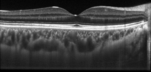
Retinal layers as seen through OCT scan
AMD
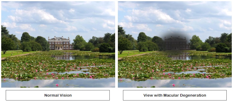
Age-related macular degeneration (AMD) is a problem with the retina. It happens when a part of the retina called the macula is damaged. With AMD you lose your central vision. You cannot see fine details, whether you are looking at something close or far. But your peripheral (side) vision will still be normal. For instance, imagine you are looking at a clock with hands. With AMD, you might see the clock’s numbers but not the hands.
There are two types of macular degeneration: dry form and wet form.
Dry form
This form is quite common. About 80% (8 out of 10) people who have AMD have the dry form. Dry AMD is when parts of the macula get thinner with age and tiny clumps of protein called drusen grow. Also, the cones and rods photo-receptors in the central retina become damaged and the central vision progressively deteriorates very slowly over a number of years.
Wet form
This form is less common but much more serious. Wet AMD is when new, abnormal blood vessels grow under the retina. These vessels may leak blood or other fluids, causing scarring of the macula. You lose vision faster with wet AMD than with dry AMD. Many people don’t realize they have AMD until their vision is very blurry. This condition can now be successfully slowed down by early intervention in order to prevent permanent loss of central vision. This is why it is important to have regular visits to your ophthalmologist.
Treating wet AMD
To help treat wet AMD, there are medications called anti-VEGF drugs. Anti-VEGF treatment helps reduce the number of abnormal blood vessels in your retina. It also slows any leaking from blood vessels. This medicine is delivered to your eye through a very slender needle.
Laser photocoagulation is another way of treating some types of wet AMD. During the procedure, a laser is used to finely cauterize ocular blood vessels
DIABETIC RETINOPATHY
People with diabetes can have an eye disease called diabetic retinopathy. This is when high blood sugar levels cause damage to blood vessels in the retina. These blood vessels can swell and leak. Or they can close, stopping blood from passing through. Sometimes abnormal new blood vessels grow on the retina. All of these changes can steal your vision. Treatment includes the use of lasers or injection of drugs that cause the swelling (from leaking blood vessels) to go resolve.
EYE FLOATERS
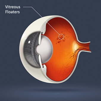
Eye floaters appear as small spots that drift through your field of vision. They may stand out when you look at something bright, like white paper or a blue sky. They might annoy you, but they shouldn’t interfere with your sight. Most floaters are small flecks of a protein called collagen. They’re part of a gel-like substance in the back of your eye called the vitreous. As you age, the protein fibers that make up the vitreous shrink down to little shreds that clump together. The shadows they cast on your retina are floaters. If you see a flash, it’s because the vitreous has pulled away from the retina. If that happens, see your eye doctor as soon as possible.
RETINAL TEARS
A retinal tear occurs when there is an abnormal attachment between the vitreous gel and the retina. In this case the vitreous gel can tug on the retina causing it to tear. If there are retinal blood vessels at the location of the retinal tear, bleeding will occur inside the eye. Retinal tears are painless. Retinal tears can cause new floaters, intermittent flashing lights, cobwebs and perhaps a shower of black dots. Some people have a lot of these symptoms while others notice hardly anything at all. Small holes and tears are usually treated with laser surgery. The procedure is performed in the office using local anesthesia. Laser is used to create tiny burns around the retinal tear. The healing that occurs after laser essentially spot-welds the retina down and prevents the tear from causing a retinal detachment. Occasionally it is not possible to place laser treatment. In this case a freezing procedure called cryopexy is used to treat the retinal tear instead.
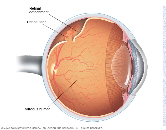
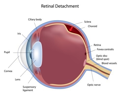
RETINAL DETACHMENT
Retinal detachment is a disorder of the eye in which the retina separates from the layer underneath. Symptoms include an increase in the number of floaters, flashes of light, and worsening of the outer part of the visual field. Also, it may be described as a curtain over part of the field of vision. If left untreated, permanent loss of vision may occur. In many cases a vitrectomy is the most commonly performed surgical operation for the treatment of retinal detachment.
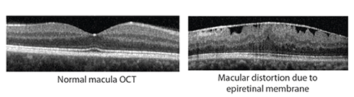
EPIRETINAL MEMBRANE
Epiretinal membranes are often related to the degeneration of the vitreous, a normal aging process inside of the eye. As the vitreous pulls away from the retina a scar-like tissue may develop. If this scar-like tissue contracts, it can cause the retina to wrinkle or pucker. The condition is mild in most cases with very few symptoms. In more severe cases the vision becomes blurred and distorted, just as one would expect a picture to appear distorted from a camera with wrinkled film. Straight lines, like doorways or telephone poles, often appear wavy. Vision loss can vary from barely noticeable to severe. For most people, vision remains stable and does not get progressively worse. Treatment is not necessary if symptoms are mild. When an epiretinal membrane causes vision loss or the symptoms start to interfere with a person’s normal daily activities then surgery is recommended. Vitrectomy surgery is the only treatment for effectively removing the membrane and improving the symptoms.