Congenital cataract

Congenital cataract is an opacity of the lens of the eye that can be present at birth.
In congenital cataract the crystalloid lens of the eye is opaque and does not allow clear vision to the baby, resulting in amblyopia.
Congenital cataract can significantly affect the vision and 24% of blindness in children worldwide is due to congenital cataract. It can affect one or both eyes of a child.
Congenital cataract can be caused several metabolic or systemic diseases, intra-uteral infections, trauma etc.
It has to be treated timely and usually treatment is surgery and long follow up.

Strabismus

In strabismus the eyes of the child are not aligned and look at different directions.

Usually one eye is the dominant eye and is looking at the direction of vision whereas the other eye converges (to the nose) or diverges (out). In some occasions the eyes alternate directions.
Strabismus can be present early, just after birth (congenital strabismus) or at a later age.
The most common strabismus is idiopathic (with no particular cause)
Often strabismus can coexist with refractive errors and correcting those can fully or partially improve the angle of strabismus.


Finally and very importantly some brain disorders can cause or coexist with strabismus and it is really crucial for the paediatric ophthalmologist to decide whether further neurological tests are necessary in a child with strabismus.

Dr. Siganos in Cyprus

We are pleased to announce Dr. Siganos’s newest collaboration with Laser Vision Nicosia eye center in Cyprus.

Laser Vision Nicosia is one of the largest and trustable eye centers in Cyprus providing comprehensive ophthalmological care services.
The complete technological infrastructure and its equipment with state-of-the-art diagnostic machines ensure better treatment of eye diseases.
The center is specially designed to meet all the requirements of safe operation, while the experienced staff monitors and cares the patient at all stages of the procedure.

Book your appointment with Dr. Siganos in Laser Vision Nicosia :

210 8210 808  (Greece)

+357 70 003937  (Cyprus)

Scleral lenses

Custom Scleral lenses are now available in our center.

Sclerals are large-diameter contact lenses that follow the shape of the cornea, whether normal or irregular.

The Scleral lenses are ideal in cases of keratoconus, high astigmatism, corneal transplantation, abnormal corneal shapes, corneal ulcer.

Also, Scleral lenses can be used as a method of treating dry eye as they protect the cornea and do not allow the lacrimal layer to evaporate.

Made of durable material, they last up to 3 years.

Due to their special structure, scleral lenses require special handling during application and removal. At our center you will be informed about the correct use and maintenance of scleral lenses. In order to select the right scleral lens, some necessary measurements of the anatomical elements of your eye and your eyesight are required. The process of choosing the right lenses for you takes about 1.5 to 2 hours.

Watch the instructional video about scleral lenses below


Book an appointment for scleral lens fitting at our call center: 210 8210 808

Keeping glaucoma under control

Glaucoma control is a state that never stops. The glaucoma patient has to visit a doctor at least once every 3 months. In case of heredity other members of your family may have to be checked.

In case you have glaucoma please follow these tips

  • Visit your eye doctor frequently. 3-4 visits per year if your glaucoma is under control.
  • Follow your medical prescription as given by your doctor, and don’t interrupt it even if your eye pressure is low.
  • Ask from your family the help and support you need.

Early diagnosis

Due to glaucoma’s non-symptomatic nature, many people are surprised when they are diagnosed with it. This is the reason you have to get examined as frequently as your doctor suggests. With early diagnosis you can prevent the excessive optic nerve damage and conserve your ocular health so you can continue to live a happy life.

Daily use of Eye Drops

-Before using your eyedrops wash your hands thoroughly. Be seated and lean your head backwards or lay down and look at the ceiling. Then follow the next steps.

-Slightly pull your lower lid using your index.

-Look up. Let a drop fall between your widely opened eyelids. Do not wipe your eyes and don’t let the vial touch your eyes, eyelids, or face.

-Close your eyes. Gently press into the inside corner of your eye (this prevents the medication to fall through your esophagus. Continue to pressure for at least 2-3 minutes.

-Repeat these steps to do the same in your other eye. Wait 2 to 5 minutes to pour ant other type of eye drops.

Side effects

There is a chance that the glaucoma eye drops or tablets may cause you some type of unwanted side effects. Contact your doctor if :

-you feel Headaches

-you feel Redness or irritation of the eyes

-The color of your iris has changed

-You feel fatigue

-You feel nausea, anorexia or gastric irritation

Immediately contact your doctor in case of

-Chest pain

-Heavy breathing

-Blurry vision

Closed angle glaucoma

The closed angle glaucoma is less common than the open angle glaucoma. Typically it causes sudden pain that, if not treated , can lead to blindness during the next 24 hours.

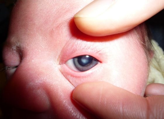
In cases of closed angle glaucoma, the iris blocks the eye’s draining system. As soon as the system is blocked the fluid that nourishes the eye cannot escape and the pressure inside the eye rises. This causes acute pain that is usually present in one eye. Other symptoms like redness, headache, nausea, pain or blurry vision can be present. In case you feel any of the above, contact with your eye doctor immediately.

  • Narrow angle glaucoma.

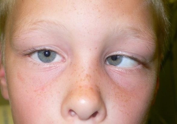
In this type of glaucoma the eye’s draining system is narrow but not blocked, fact that causes a gradual increase of the eye pressure, and makes the eye prone to closed angle glaucoma. Usually both eyes have narrow draining systems. Due to the lack of symptoms you have to get your eyes regularly checked for any changes, that may be caused by narrow angle glaucoma.

  • Treating closed angle glaucoma

To treat the closed angle glaucoma the intraocular pressure has to immediately drop, so the optic nerve damage and the vision loss can be avoided. This includes eye drops combined with laser therapy.

  • Medication treatment

Prescribing medication is ideal in cases where the intraocular pressure has to be lowered. Eye drops help at lowering the fluid production and improve the eye’s fluid draining system. Tablets or intravenous medication also help the body to reject the extra fluid.

  • Iridectomy

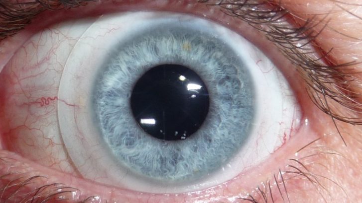
This laser assisted method is used to bypass the eye’s blocked draining system. A small hole is opened in the periphery of the iris so the fluid can travel from the posterior to the anterior chamber of the eye. This type of therapy is also used as a preventive method for glaucoma in patients prone to glaucoma.

  • During Laser treatment

The whole process lasts for a few minutes

-Anesthetic eye drops are going to be placed at the affected eye

-The patient sits in front of the laser apparatus and the doctor properly focuses the laser beam on the iris

-During the procedure there is a chance of slight inconvenience or eye pain

-Patients vision may be blurry for the next few days

-The doctor will indicate when is the right time to go back to everyday activities

-The day after your treatment the doctor will measure your intraocular pressure and give you post operative instructions.

Open angle glaucoma

Primary open angle glaucoma is a subset of the glaucoma defined by an open, normal appearing anterior chamber angle and raised intraocular pressure (IOP), with no other underlying disease. It is the most common type of glaucoma and most patients do not realize they have it until the damage is extended. Open angle glaucoma gradually gets worst through time and it usually affects both eyes. Treating open angle glaucoma includes eye drop medication, tablets and surgical or laser assisted procedures.

  • How it works

The name of this type of glaucoma indicates that the angle the iris and the lens form is open, and the aqueous humor can pass freely to the trabecular meshwork, the “drain” of the eye. But in this type of glaucoma the trabecular meshwork may either be blocked, or disfunction in any way, leading to increased fluid concentration and the rise of the intraocular pressure.

  • Symptoms

This type of glaucoma rarely causes pain or any other symptoms. The peripheral vision fades in such a slow rhythm that sometimes the central vision is affected without a warning. The only way of glaucoma prevention is the early diagnosis.

  • Treating open angle glaucoma

Treating glaucoma can include eye drops or tablets that help the IOP to drop to normal levels. It is really important to follow your doctor’s orders about the dosage and the frequency of your medication, and never skip it even if your IOP is under control.

  • Useful advice on glaucoma medication

-The patient has to know the name, the purpose and the dosage of their medication

-The doctor has to be fully informed about other medication or supplements the patient is receiving

-In case of side effects contact your doctor

-The patient should not stop or interrupt their medication without doctor’s approve.

  • Following advice

For better results the patient has to receive their medication everyday at the same time. To achieve this, follow the next steps.

-Place your medication always at the same spot.

-Use an alarm clock to remind you the time of your medication.

-Make sure you take your medication with you when traveling

-In case you forget to receive a dosage, take it as soon a s you remember. Then continue normally your prescription.

Glaucoma screening tests

Vision loss caused by glaucoma cannot be recovered. This is why early diagnosis and treatment can prevent intensive damage of the optic nerves and vision loss. The first step is a full eye check up.

  • Medical History

During the exam your doctor will ask

If you have any relatives with glaucoma

If there is any medication you receive

If you had health issues such us heart problems, hypertension or diabetes

If you ever had an eye injury

  • Measuring the IOP

The pressure that builds up inside the eye is called intraocular pressure (IOP). The High pressure is usually a sign of glaucoma. To measure to the IOP your doctor will use an instrument called tonometer. This device will deliver a small air puff on your eye, or may touch it after your eye has been numbed with eye drops.


  • Gonioscopy

The doctor will also examine your eyes with a special lens called gonioscopy lens. This lens allows your doctor to evaluate the internal drainage system of the eye, also referred to as the anterior chamber angle. The “angle” is where the cornea and the iris meet. This is the location where fluid inside the eye (aqueous humor) drains out of the eye and into the venous system.

  • Optic disk examination

Your doctor will examine the optic disks of your eyes for any signs of damage or nerve fiber loss. Special eye drops that dilate your pupils will be used before this examination, so the doctor can have full visual access to the back of your eye. Then the doctor will use special magnifying lenses and a lighting instrument to examine the optic disk.

  • Visual Fields test

The visual field test is a subjective measure of central and peripheral vision, or “side vision,” and is used by your doctor to diagnose, determine the severity of, and monitor your glaucoma. The most common visual field test uses a light spot that is repeatedly presented in different areas of your peripheral vision. You will have to click a small button every time you perceive a beam of light. The results of the examination are analyzed with the help of a computer, and they are printed on point.

Eye with glaucoma

In glaucoma the draining holes of the eye are narrow or blocked, causing the aqueous humor to concentrate inside the eye rising the Intraocular pressure (IOP).

High IOP levels destroy the optic nerve fibers and cause peripheral view loss. The higher the IOP rises the more nerve fibers get destroyed. When the nerve fibers are lost the light cannot reach the visual cortex, so there are black spots where an image is supposed to be seen.

All of the optical nerve fibers are gathered in the back of the eye and form the optic nerve which during fundus examination looks like an orange disk. When the optic fibers go thinner, this disk gets empty. This is how your doctor can easily make a first estimation about whether or not you suffer from glaucoma.


1 2